Sneak Thief of Vision
The role of Glaucoma as a cause of blindness has been known since the 19th century. But it can be controlled if it is diagnosed and treated early
By Dr Geetika Khurana
Glaucoma is defined as chronic and progressive optic neuropathy, associated with visual field loss, which may or may not be associated with increase in intra ocular pressure (IOP).
A major risk factor for glaucoma is increased pressure in the eye. Normal IOP is in the range of 10 to 21 mmHg. The increased pressure can damage the optic nerve, which transmits images to the brain. If damage to the optic nerve from high eye pressure continues, glaucoma can cause permanent loss of vision. Without treatment, glaucoma can cause total blindness permanently, within a few years. However, there is no specific level of elevated eye pressure that definitely leads to glaucoma. Conversely, there is no lower level of IOP that will absolutely eliminate a person’s risk of developing glaucoma. Because most people with glaucoma have no early symptoms or pain from this increased pressure, it is important to consult eye doctor regularly so that glaucoma can be diagnosed and treated before permanent visual loss occurs.
Pathophysiology
Glaucoma is usually caused when pressure in one’s eye increases. This can happen when intraocular fluid isn’t circulating normally in the anterior part of the eye. Normally, this fluid called aqueous humour flows out of the eye through a mesh-like channel called trabecular meshwork. If this channel becomes blocked, fluid builds up, causing glaucoma. The direct cause of this blockage in primary glaucoma is unknown, but it is found to have a genetic predisposition. Less common causes of glaucoma include a blunt or chemical injury to the eye, blockage of retinal blood vessels in the eye, inflammatory conditions of the eye, and occasionally eye surgery to correct another condition. Glaucoma usually occurs in both eyes, but it may involve each eye to a different extent.
Types of Glaucoma
There are two main types of glaucoma:
1. Open-angle glaucoma
This is the most common type of glaucoma. The structures of the eye appear normal, but fluid in the eye does not flow properly through the trabecular meshwork.
2. Angle-closure glaucoma
It can be acute or chronic angle-closure or narrow-angle glaucoma. This type of glaucoma is less common in the West than in Asia. Poor drainage is caused because the angle between the iris and the cornea is too narrow and is physically blocked by the iris. This condition leads to a sudden buildup of pressure in the eye.
Although “normal” eye pressure is considered a measurement less than 21 mmHg, this can be misleading. Some people have a type of glaucoma called normal-tension, or low-tension glaucoma. Their eye pressure is consistently below 21 mmHg, but optic nerve damage and loss of vision still occur. People with normal-tension glaucoma are usually treated in the same way as people who have open-angle glaucoma.
Congenital glaucoma is a rare type of glaucoma that develops in infants and young children and can be inherited. While less common than the other types of glaucoma, this condition can be devastating, often resulting in blindness if not diagnosed and treated early.
Secondary glaucoma results from another eye condition or disease. For example, someone who has had an eye injury, someone who is on long-term steroid therapy or someone who has a tumour may develop secondary glaucoma. The most common forms of secondary glaucoma are: pseudoexfoliative glaucoma, pigmentary glaucoma, and neovascular glaucoma.
Some people have normal eye pressure but their optic nerve or visual field looks suspicious for glaucoma. These patients are known as glaucoma suspects. They must be watched carefully because some may eventually develop definite glaucoma and need treatment.
Other people have an eye pressure that is higher than normal, but they do not have other signs of glaucoma, such as optic nerve damage or blank spots that show up in their peripheral (side) vision when tested. This condition is called ocular hypertension. Individuals with ocular hypertension are at higher risk for developing glaucoma compared to people with lower, or average, eye pressure. Just like people with glaucoma, people with ocular hypertension need to be closely monitored by an ophthalmologist to ensure they receive appropriate treatment.
Risk Factors
• Age above 40 years
• African-American, Irish, Russian, Japanese, Hispanic, Inuit, or Scandinavian descent
• Family history of glaucoma
• Diabetes
• Long term use of topical or oral steroid
• Trauma
Symptoms
There are usually few or no symptoms of glaucoma. The first sign of glaucoma is often the loss of peripheral or side vision, which can go unnoticed until late into the disease. This is why glaucoma is often called the “sneak thief of vision”.
Detecting glaucoma early is one reason one should have a complete eye test with an eye specialist every one to two years, especially after age of 40 years. Occasionally, intraocular pressure can rise to severe levels which can cause sudden eye pain, headache, blurred vision, or the appearance of halos around lights.
Diagnosis
To diagnose glaucoma, an ophthalmologist will test the vision and examine your eyes through dilated pupils. The eye exam typically focuses on the optic nerve, which has a particular appearance in glaucoma. In fact, photographs of the optic nerve can also be helpful to follow over time as the optic nerve appearance changes with the progression of the disease. The doctor will also perform a procedure called tonometry to check for eye pressure, and a visual field test, if necessary, to determine if there is loss of peripheral vision. Glaucoma tests are painless and take very little time.
Treatment
Various treatment modalities for glaucoma include:
• Topical therapy: These either reduce the formation of aqueous humor or increase its outflow. Side effects of anti-glaucoma drops may include allergy, redness of the eyes, brief stinging, blurred vision, and irritated eyes. Some glaucoma drugs (beta blockers) may affect the heart and lungs.
• Laser procedures for glaucoma. Laser surgery for glaucoma slightly increases the outflow of the fluid from the eye in open-angle glaucoma or eliminates fluid blockage in angle-closure glaucoma. Types of laser surgery for glaucoma include trabeculoplasty, in which a laser is used to pull open the trabecular meshwork drainage area; iridotomy, in which a tiny hole is made in the iris, allowing the fluid to flow more freely; and cyclophotocoagulation, in which a laser beam treats areas of the middle layer of the eye, reducing the production of fluid.
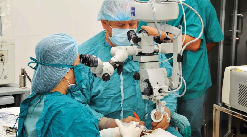
• Microsurgery for glaucoma. In an operation called a trabeculectomy, a new channel is created to drain the fluid, thereby reducing intraocular pressure that causes glaucoma. Implantation of drainage devices is also required in certain cases. Complications of microsurgery for glaucoma include failure, temporary or permanent loss of vision, bleeding and infection.
Glaucoma cannot be prevented but it can be controlled if it is diagnosed and treated early. Loss of vision caused by glaucoma is irreversible. However, successfully lowering eye pressure can help prevent further visual loss from glaucoma. Most people with glaucoma do not go blind if they follow their treatment plan and have regular eye tests by eye specialists.
(The author is Senior Resident, Army College of Medical Sciences & Base Hospital, New Delhi)

